
Laser Surgery
 Laser surgery of the retina can be used to treat a wide range of retinal conditions through minimally invasive techniques that produce long-lasting results. These diseases can often lead to serious complications such as hemorrhaging and blindness. Lasers have been used to treat eye diseases for more than 30 years and produce effective results with no scalpal or needles.
Laser surgery can be used to treat diabetic retinopathy, retinal vein occlusions, age-related macular degeneration, retinal detachments and more. Depending on the patient’s condition, the laser may be used to seal leaking blood vessels, repair tears, remove newly formed blood vessels or destroy tumors. These procedures are performed in the doctor’s office and require only anesthetic eye drops to numb the area prior to treatment. Laser treatment usually takes less than 30 minutes to perform, and patients can go home immediately following surgery. Most patients return to work and other normal activities the next day.
Several laser treatments may be needed in order to achieve optimal results and to help manage chronic retinal conditions. Results may take a few weeks or months to become noticeable, so it is important to see your doctor for follow-up appointments in order to ensure that you receive the best possible results.
Focal Photocoagulation
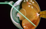
Retinal laser photocoagulation is a minimally invasive procedure used to seal or destroy leaking blood vessels in the retina that lead to serious retinal conditions such as diabetic retinopathy and macular edema. This procedure can also seal retinal tears and destroy abnormal tissue found in the back of the eye.
Focal laser coagulation may be recommended for patients with clinically significant macular edema (CSME) – swelling of the central retina, called the macula. The laser coagulates, or dries up, the fluid that is causing the swelling. During focal photocoagulation, laser burns are made on the retina to target leaking blood vessels. While it cannot restore vision that has already been lost, it can reduce the risk of vision loss.
This procedure is performed with a local or topical anesthetic on an outpatient basis. Patients will need someone to drive them home after the procedure, since the pupils will be dilated for several hours. Your vision may also be blurry and you may experience mild pain for a day or two after the procedure. You can resume normal activities immediately.
Laser photocoagulation carries certain risks, since it involves burning and destroying part of the retina. Patients may experience a mild loss of central vision, reduced night vision and a decreased ability to focus. However, the potential vision loss caused by this procedure is far less than the severe vision loss that can occur as a result of retinal conditions like diabetic retinopathy.
Pan Retinal Photocoagulation
 Pan retinal photocoagulation is a more extensive variation on focal photocoagulation. It is used to treat the entire area to slow the growth of new abnormal vessels. This form of treatment is reserved for more serious cases of retinopathy.
During pan retinal photocoagulation, a laser is applied to all portions of the retina with blood vessel damage and tissue that has not received enough oxygen. The blood vessels will shrink, and new ones are not created. This can lower your risk of further vision loss due to a retinal detachment or vitreous hemorrhage.
Micro-Pulse Technology
A newer form of pan retinal photocoagulation, micro-pulse technology allows the doctor to control the timing and intensity of the laser pulses in order to curtail treatment time and maximize precision. These short pulses are applied to the abnormal blood vessels mere milliseconds apart, providing a greater level of safety to the patient because it can be more precisely targeted and reduce the likelihood of damage to the nearby epithelium.
Photodynamic Therapy
 Photodynamic therapy is used to treat a complication of wet macular degeneration in which leaks form beneath the retina. These leaks occur in a structure in the eye called the choroidal neovascular membrane, or CNVM. During photodynamic therapy, dye is injected and a narrow-wavelength laser beam is focused on it for about 90 seconds. The dye absorbs the energy and slows or stops leakage by creating blood clots and stopping abnormal blood vessels from growing. Following treatment, patients should avoid direct exposure to sunlight for several days. Patients most likely to benefit from treatment will have newly onset macular degeneration and no scarring. Vision stabilization is maximized with a series of treatments over one to two years.